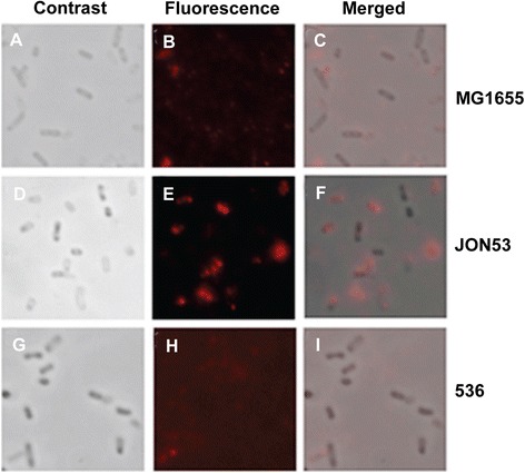
Figure 3.

Immunofluorescence microscopy detection of ClyA on bacterial cells. Analyses were done with the E. coli K-12 strain MG1655 (A, B C), the clyA + UPEC derivative JON53 (D, E, F) and the parental UPEC strain 536 (G, H, I). Panels A, D, G show images obtained by by phase contrast microscopy. Panels B, E, H show images obtained from immunofluorescence analysis using polyclonal ClyA antiserum and AlexaFluor 555 –conjugated secondary antibody to enable visualization of ClyA as a red fluorescence signal. Panels C, F, I show the merged images.
