Figure 3.

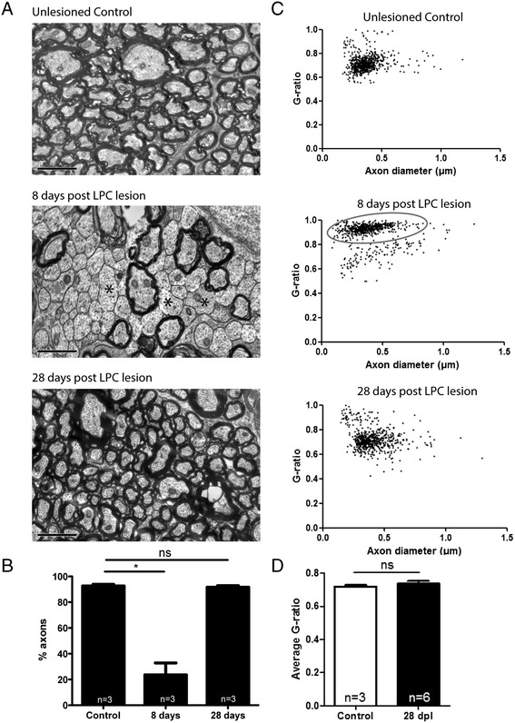
In young zebrafish the proportion of myelinated axons and myelin thickness is fully restored after demyelination. (A) In lesioned optic nerve, electron microscopic analysis shows many demyelinated axons at 8 dpl (asterisks) and complete remyelination at 28 dpl compared to unlesioned controls. (B) At 8 dpl the number of myelinated axons in the lesion area is significantly reduced compared to unlesioned controls (p < 0.05, n = 3 fish, Kruskal Wallis test, Dunn’s post-test), while there is no difference between remyelinated and control optic nerves. (C) Graphs of G-ratio versus axon diameter show similar patterns at 28 dpl and in unlesioned controls, while most axons at 8 dpl have a higher G-ratio (less myelin). (D) Average G-ratio between unlesioned and remyelinated optic nerve is not different, indicating remyelination with normal thickness myelin (p > 0.05, n = 3-6 fish, Mann Whitney U-test). Mean ± SEM. Scale bar: A = 1 μm.
