
Figure 3. Arsenic trioxide and ascorbic acid induced cytotoxicity in CLL cells is dependent on activation of caspase.
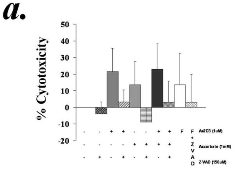
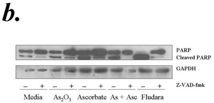
Purified B-lymphocytes from CLL patients (1×10⁶/ml media) were incubated with 1μM arsenic and 1mM ascorbic acid either alone or in combination in the presence or absence of z-VAD-fmk (150uM) for 24 hours. The cells were stained with Annexin-V-FITC and propidium iodide and analyzed by flow cytometry for apoptotic cells (panel a). The data shown represent % Annexin-V+/PI+ cells ± SD.(n=3). Panel B shows Westernblot analysis of the lysates from one of the above experiments was assessed for incleaved and cleaved PARP. Fludarabin is used as a control in these studies.