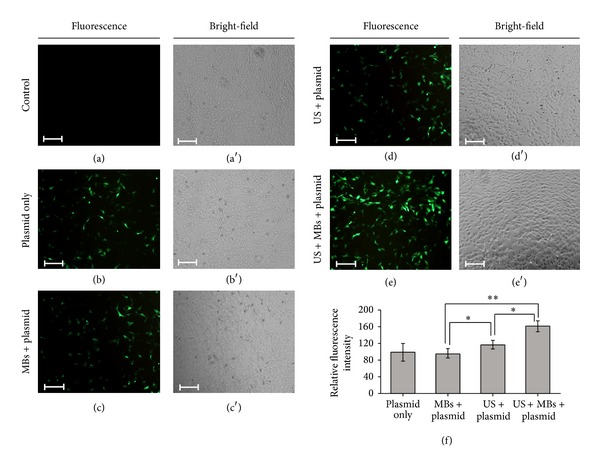
Figure 5.

The transfection efficiency of HEI-OC1 cells with plasmid DNA by using a combination of US and MBs. (a, a′) Fluorescence and bright-field images of living cells in the control groups, (b, b′) plasmid only, (c, c′) MBs with plasmid, (d, d′) US with plasmid, and (e, e′) US with MBs and plasmid. (f) Fluorescence intensities in the five groups quantified and normalized relative to the control group. Scale bar = 200 μm. Results are expressed as mean ± standard deviation with n = 5 for each bar. *indicates P < 0.05; **indicates P < 0.01.
