Figure 1.
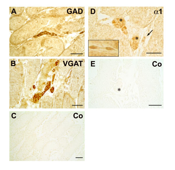
Leydig cells in postnatal rat testis possess GAD, VGAT and GABAA-α1. Immunohistochemical localization of GAD, VGAT and GABAA-α1 in the testis of five days old rats. Fetal Leydig cells with typical rounded morphology are located in clusters between the seminiferous tubules and are immunopositive for GAD (A) and VGAT (B). No reaction was observed in sections incubated with buffer (data not shown) or non-immune rabbit serum (C) instead of the primary antibody. The GABAA receptor subunit α1 (D) is also immunolocalized to clustered fetal Leydig cells (*). However other interstitial cells with a spindle-shaped appearance also exhibit specific immunoreactivity against GABAA-α1 (D, →). The insert panel (D) also depicts magnified spindle-shaped interstitial cells of another section, which are immunopositive for GABAA-α1. No reaction was observed in sections incubated with buffer (data not shown) or non-immune rabbit serum (E) instead of the primary antibody. Bars: 50 μm.

