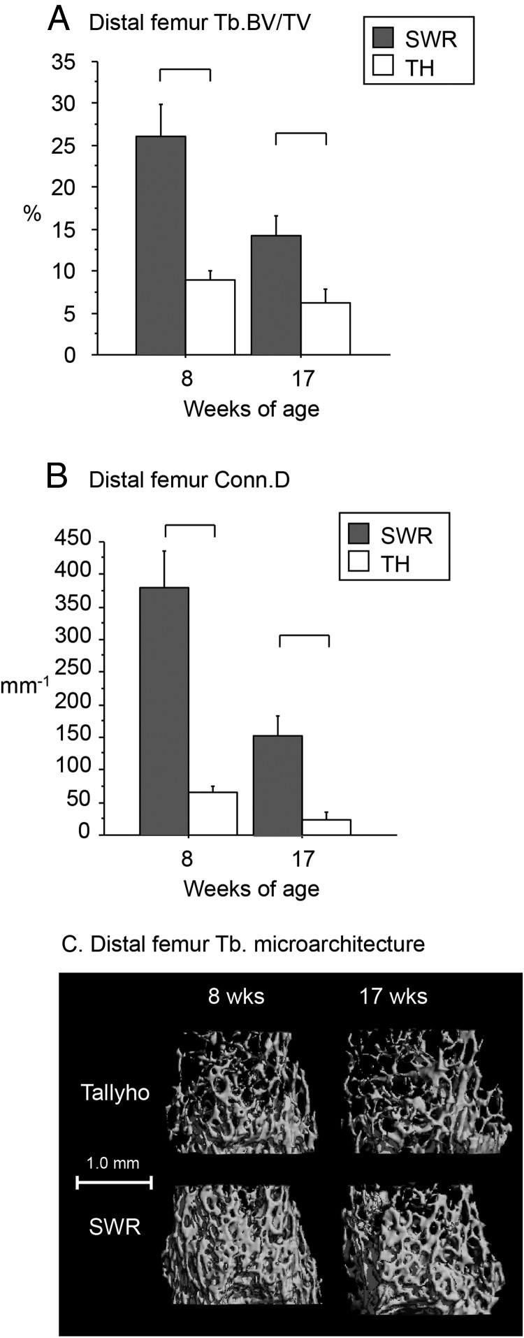
Figure 2.
In the distal femur, trabecular BV/TV (A; percentage) and Conn.D (B; per millimeter) were markedly lower in Tallyho vs SWR at 8 and 17 weeks of age. C, Overall, Tallyho exhibited severely impaired trabecular microarchitecture at 8 and 17 weeks of age. Significant differences (P < .05) are indicated by brackets.

