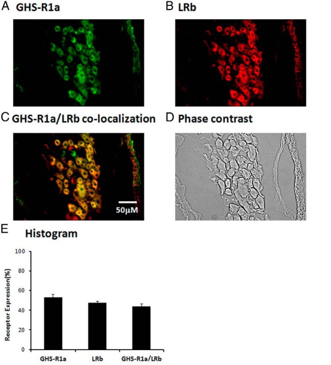
Figure 1.
Immunostaining of rat NG neurons demonstrates colocalization of GHS-R1a and LRb. A, Representative photomicrograph of a normal rat NG stained for GHS-R1a shows 53.1 ± 3% of NG neurons stained positive for GHS-R1a. B, 47.2 ± 2% of NG neurons stained positive for LRb. C, 91 ± 3% of LRb-bearing NG neurons contained GHS-R1a. D, Phase contrast image of a normal rat NG to demonstrate the number of cells. E, Histogram shows the distribution of GHS-R1a and LRb expression. The data are representative of three independent experiments.

