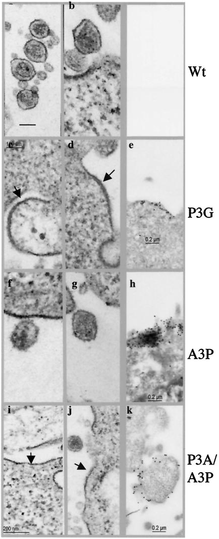
FIG. 5.
Phenotypes of late domain mutants by electron microscopy. HeLa cells were transfected with the indicated pCMVHT-1 constructs. Arrows indicate a thickening of the plasma membrane due to the retention of viral proteins. (a to d, f, g, i, and j) TEM images; (e, h, and k) IEM images. Staining was done with a monoclonal anti-MA antibody followed by anti-mouse immunoglobulin G-gold (5-nm diameter).

