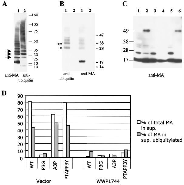
FIG. 8.
MA is ubiquitinated in a WWP1-dependent fashion. (A) Immunoblot analysis of density-separated virus from HTLV-1-infected MT2 cell line with an anti-MA antibody (left) and an anti-ubiquitin antibody (right). Arrows indicate comigrating bands. (B) Pelleted supernatants from 293T cell cultures transfected with pCMVHT1 (lanes 1) or left untransfected (lanes 2) were denatured and immunoprecipitated with a rabbit anti-MA antibody (see Materials and Methods). The right panel was probed with a mouse monoclonal anti-ubiquitin antibody. After stripping, the blot was reprobed with a mouse monoclonal anti-MA antibody (left). The positions of molecular weight markers are given on the right. * and **, predicted positions for mono- and di-ubiquitinated MA, respectively. (C) Immunoblot analysis with 2 ng of the respective matrix protein (according to ELISA) loaded per lane. The mutants used were pCMVHT (lane 1), pCMVHT-P3G (lane 2), pCMVHT-A3P (lane 3), pCMVHT-P3AA3P (lane 4), pCMVHT-PTAP/P3Y (lane 5), and pCMVHT-PTAP/P3G (lane 6). The blot was probed with an anti-MA monoclonal antibody and developed with ECL+ and autoradiography. (D) 293T cells were transfected with 2 μg of the indicated pCMVHT1 constructs plus 15 μg of pUCHR or pUCHR-WWP1744. The fractions of MA in the supernatants were determined by ELISA and are shown as open bars. The fractions of MA from the supernatants that were ubiquitinated are shown as solid bars and were established as described in the text.

