Figure 5.
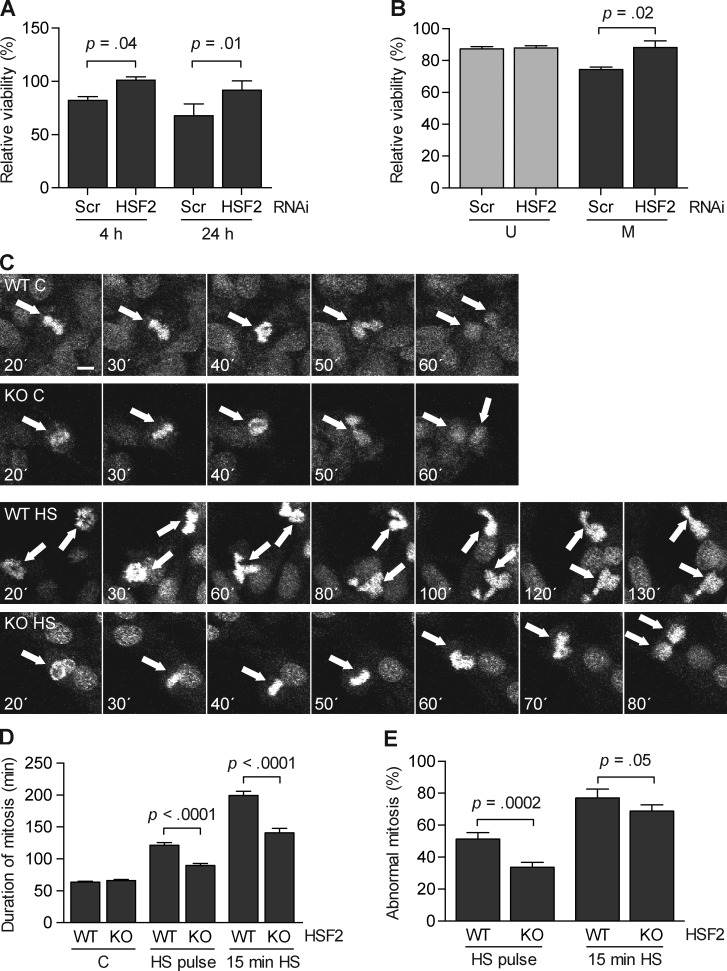
Diminished HSF2 levels promote cell survival after heat shock in mitosis. (A) K562 cells transfected with the indicated shRNA constructs were synchronized into mitosis by a thymidine-nocodazole block and exposed to heat shock (30 min at 42°C). Cells were counted in triplicate after 4 h and 24 h of recovery at 37°C. The number of cells was related to their untreated counterparts (n = 4). (B) HeLa cells transfected as in A were left unsynchronized (U) or synchronized into mitosis and collected by mitotic shake-off (M) before being heat-treated (30 min at 43°C) or left untreated. Survival was measured by an MTS assay after a 2-h recovery at 37°C. Values from the heat-treated samples were related to their respective control (n = 4). (C) Selected still frames from time-lapse microscopy of DRAQ5-stained HSF2 WT and HSF2 KO MEFs that were either left untreated (C) or subjected to a pulse of heat shock at 43°C (HS; for details see Materials and methods). Time is given as minutes after heat shock. Arrows denote mitotic cells. Bar, 10 µm. See Videos 1–4. (D) Duration of mitosis was measured from nuclear envelope breakdown to cytokinesis in untreated (C) or heat-treated (pulse at 43°C or 15 min at 43°C) HSF2 WT and HSF2 KO MEFs. At least 20 cells per treatment from four individual experiments were monitored. (E) Ratio of abnormal mitosis in cells from D. Abnormal mitosis was measured as the percentage of cells with defects in chromosome segregation, such as daughter cells forming micronuclei, chromatin decondensing without division, formation of more than two daughter cells, or apoptosis. All values represent mean + SEM (error bars).

