
Figure 2.
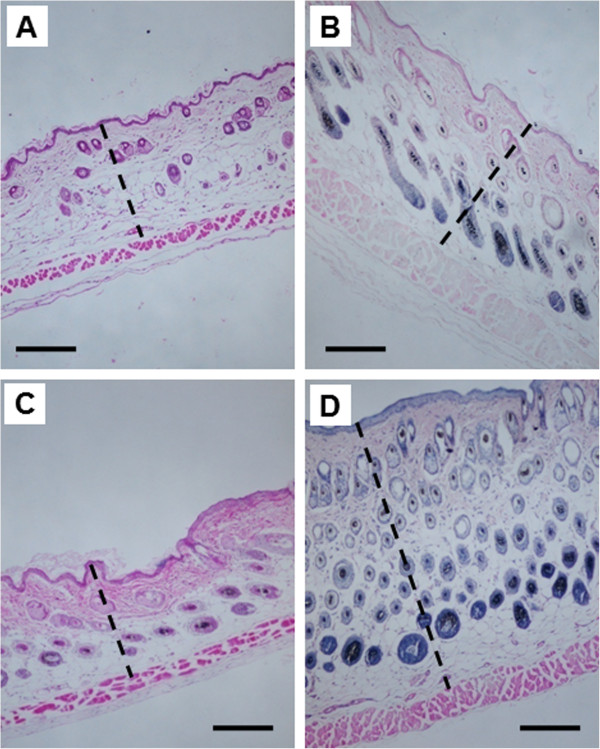
Skin thickness in Nrf2 WT versus Nrf2 KO mice after UVB exposure. H&E staining was performed in skin samples 8 days after treatment with a single dose of UVB (300 mJ/cm2). WT mice were treated (A) without UVB or (B) with UVB. KO mice were treated (C) without UVB or (D) with UVB. The horizontal line represents 200 μm, and the broken lines indicate the skin thickness. Original magnification, ×400. The Nrf2 KO mice exhibited more edema and inflammatory changes, such as increased skin thickness, whereas the Nrf2 WT mice demonstrated fewer changes; these data correlated with the ear punch weights (Figure 1) and other biomarkers (Figures 3, 4, 5).