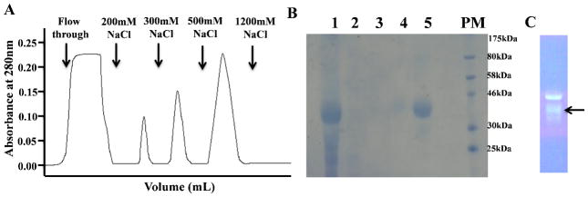
Fig 3.

Purification of hIL-12 using heparin Sepharose chromatography. (A) Fractionation of proteins, secreted in the hollow fiber culture medium, on heparin Sepharose. (B ) SDS-PAGE of the different fractions, eluted under a NaCl gradient, as detected by Coomassie blue staining. Lane 1 - Cell Culture Supernatant; Lane 2 - Flow through (100 mM NaCl); Lane 3 – 200mM NaCl; Lane 4 – 300mM NaCl; Lane 5 – 500mM NaCl; PM-Protein Marker. (C) Lane showing the purified hIL-12 from 500mM NaCl fraction stained with ProQ emerald green stain and a distinct p35 subunit band(s) is highlighted by an arrow.
