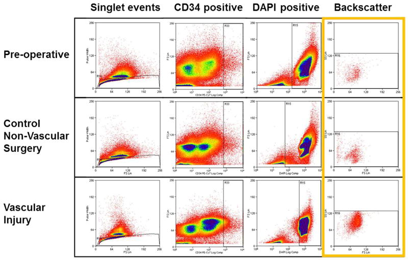
Fig. 2.
Flow analysis of sheep for CD34-positive cells. Blood specimens drawn preoperatively, or from the first postoperative day after nonvascular or vascular surgery were compared. A total of 300,000 events from peripheral blood specimens were gated sequentially for singlet events, CD34 positivity, DAPI positivity, and finally backscattered to reveal final population of cells highly expressing CD34. Increased numbers of CD34-positive cells can be seen after vascular injury (bottom right).

