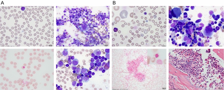
Figure 1.

Comparison of peripheral blood and bone marrow morphology at 2 months (A) and 4 months of age (B). In panel A: Top left: Peripheral blood showed normocytic RBCs without anisopoikiocytosis. A very rare agranular large platelet is seen (MPV 10.3). Top right: Bone marrow aspirate showed trilineage hematopoiesis with marked erythroid hypoplasia. The megakaryocytes appeared unremarkable. Bottom left: Iron stain on the bone marrow aspirate showed a rare ringed sideroblast. Bottom right: Occasional early stage myeloid cells contained small cytoplasmic vacuoles. In panel B: Top left: Peripheral blood showed normocytic anemia with marked anisopoikilocytosis. The insert showed the presence of one of the many RBCs with coarse basophilic stippling. There were many giant agranular platelets (MPV 12.9). Top right: The bone marrow aspirate showed moderate dyserythropoiesis with increased monolocated/hypolobated megakaryocytes with eccentrically located nucleus (insert). Bottom left: The bone marrow showed increased iron storage for age with rare ringed sideroblasts (insert). Bottom right: The bone marrow core biopsy showed the presence of the small megakaryocytes with crescent shaped eccentrically located nuclei.
