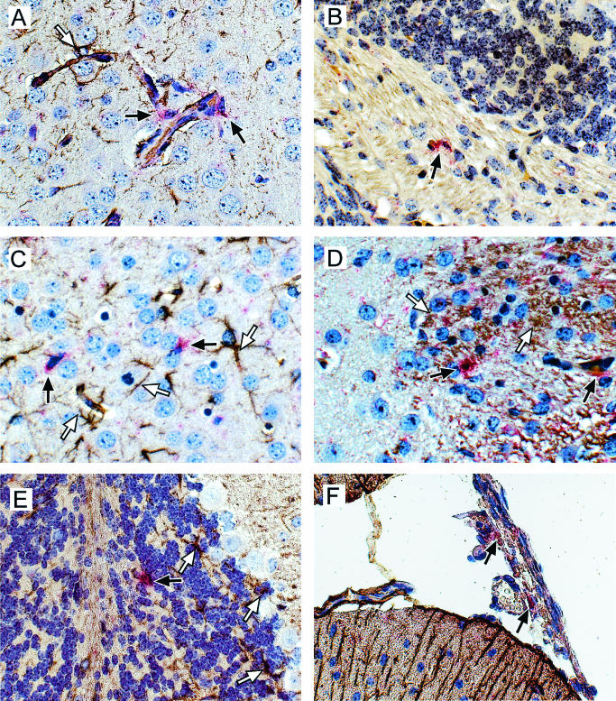
FIG. 6.
In situ hybridization and immunohistochemistry analysis of MCP-5 expression in Fr98-infected IRW mice (A to D) or Fr98-NSC-inoculated BALB/c mice (E and F). Sections were hybridized with DIG-labeled antisense RNA for MCP-5 and developed using Fast Red stain (red cytoplasm color, black arrows). No Fast Red-positive cells were observed in mock-infected mice for any of the above chemokines (data not shown). Sections were then incubated with anti-GFAP (A, C, E, and F), no antibody (B), or anti-gp70 virus envelope (D) and developed with DAB (brown/black positive color, white arrows). All sections were counterstained with hematoxylin. Photographs were taken using a digital camera. Magnification, ×40.

