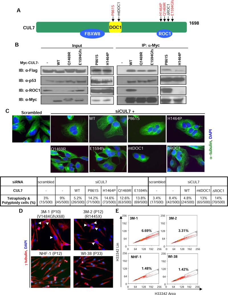
Figure 3. 3M derived mutations in CUL7 disrupt its function in regulating microtubule dynamics and maintaining genome integrity.
(A) Schematic representation of human CUL7 protein. 3M patient derived mutations are indicated in red. The mutations targeting two functional domains, DOC1 and ROC1 binding, are also indicated.
(B) 293T cells were cotransfected with plasmids expressing indicated proteins. Two days after transfection, expression and association between CUL7 with FBXW8, p53 and ROC1 was determined by western or IP-western analyses.
(C) U2OS cells were transduced with retrovirus encoding siRNA resistant, Myc-tagged wild-type or various CUL7 mutants. Cells were then transfected with scrambled or CUL7 siRNA, followed by α-tubulin and DAPI staining. Images are representative views of each sample. Five hundred cells from each group were scored for polyploidy. See also Figure S3.
(D, E) Normal human forehead fibroblasts NHF1, normal human lung fibroblasts WI38, and skin fibroblasts derived from two 3M patients bearing CUL7 mutations were stained with α-tubulin (red) and DAPI (blue). Five hundred cells of each group were counted to score tetraploid cells (D), or stained with Hoechst 33342 and then subjected to FACS analysis to quantify polyploidy cells (E).

