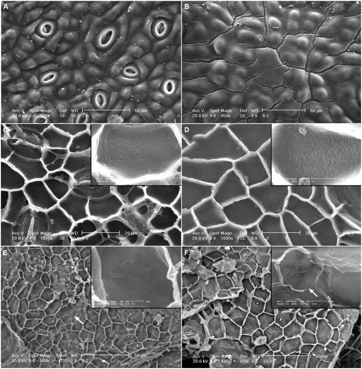
FIGURE 4.
SEM micrographs of the adaxial leaf cuticles of E. camaldulensis and E. globulus after successive extractions. Outer side of E. camaldulensis (A) and E. globulus (B) SE cuticles. Inner side and detail of E. camaldulensis (C) and E. globulus (D) SE cuticles. Inner side and detail of E. camaldulensis (E) and E. globulus (F) SED residues. SE: Soxhlet extracted, SED: Soxhlet extracted, and depolymerized. Bars in insert images: 5 μm (C,D,F), 2 μm (E). Arrows indicate areas were the anticlinal cuticle was not visible (E,F).

