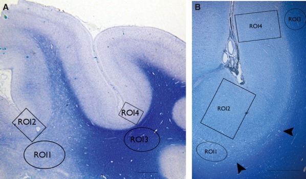
Figure 1.

Low power views of myelin stained sections (LFB) form two cases of FCD type IIB illustrating the regions of interest (ROIs) used for the analysis. (A) The white matter pallor extends from the depth of sulcus deep to the white matter, whereas in (B) only the immediate subcortical zone, that of the U-fibers shows pallor that forms a band running along the bottom of the cortex (arrowheads) and the overlying cortex shows excess myelination. The ROI indicated are ROI 1 subcortical white matter (WM) in region of dysplasia, ROI2 dysplastic cortex (full thickness) overlying ROI1, ROI 3 normal WM in adjacent cortex, ROI4 normal cortex (full thickness) overlying ROI 3. (The ROI shown here provide an approximation of the size of the freehand drawn ROI on the immunostained sections.) The scale bars in A = 800 and B = 1,500 μm.
