Figure 2.

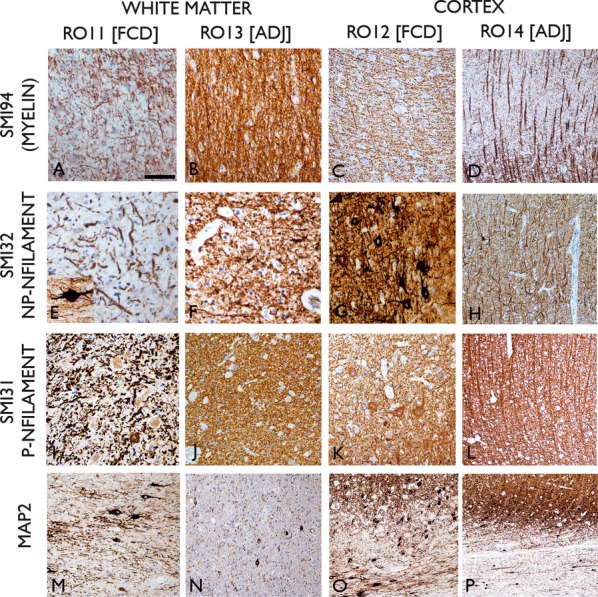
Immunohistochemistry for myelin basic protein (SMI94; A–D), nonphosphorylated neurofilament (NP-NFilament SMI32; E–H), phosphorylated neurofilament (P-Nfilament SMI31; I–L) and Map2 (microtubule associated protein) in ROI1 (FCD WM), ROI3 (normal WM), ROI2 (FCD cortex), and ROI4 (normal cortex). Reduction of number of processes was noted in ROI1 with SMI31,32, & 94 antibodies with thick, tortuous fibres present, particularly in SMI32. Inset in (E) shows a dysmorphic neuron in the immediate subcortical region with thick bipolar processes running horizontally to the cortex. In ROI3 (B, F, J) normal density and size of axons were seen with all antibodies. In the dysplastic cortex, prominent horizontal fibers were seen with SMI94 (C), obscuring the normal radial orientation observed in normal cortex (D). Similarly in neurofilament stains, disorganized axonal and dendritic processes were seen in the dysplasia (G, K) relative to the radial organized patterns of normal cortex (H, L). In Map2 stained sections in the WM of the region of dysplasia (M), dysmorphic neurons and dendrites were present compared to infrequent, small white matter neurons and fine dendrites in adjacent normal WM (N). In the region of dysplasia (O) Map2 staining highlights the ill-defined border between the gray and white matter interface with numerous unstained balloon cells and prominent horizontal neurons in the subcortical zone. In the adjacent cortex, sharper demarcation of cortex and white matter is observed (P). ROI, Region of interest; FCD, Focal cortical dysplasia; WM, white matter; ADJ, adjacent normal cortex. Bar = 60 microns in A to N and 140 microns in O & P.
