
Figure 3.
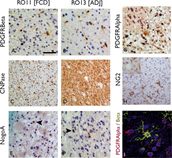
Immunohistochemistry for oligodendroglial (OL) and precursor cell types (OPC). Comparison of ROI1 (white matter in the region of dysplasia [A, C, E]) with ROI3 (adjacent white matter [B, D, F]) positive labeling of cells with PDGFRβ (A, B) CNPase (C, D) and NogoA (E, F) are seen in both ROI. With PDGFRβ, small round cells were labeled with fine branching processes, compatible with the described morphology of OPC, and were visible in both ROI; with CNPase, labeling of small OL in addition to fibers was noted with a marked reduction in the labeling of fibers in ROI1 (C) compared to ROI3 (D). NogoA labeled infrequent small OL cells in all ROI with a small, peripheral rim of cytoplasmic labeling. (G) PDGFRα also showed positive round cells in ROI1 and (inset) ROI3. (H) NG2 labeled cells with similar morphology, with fine branching process in ROI3 and in (inset) ROI1 near to an unlabeled balloon cell. (I) Confocal microscopy confirmed overlap of labeling of PDGFRα and β in cells with multipolar morphology. Bar = 15 microns (A, B, E, F, G, H, I [including insets]) and 35 microns (C, D).