
Figure 6.
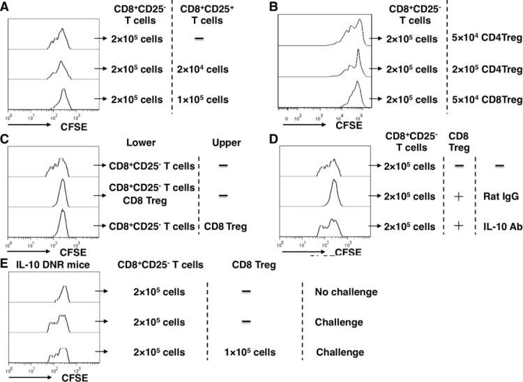
CD8+CD25− T-cell proliferation was inhibited by CD8+ Treg cells through IL-10 in vitro. (A) CD8+CD25+ T cells, CD8+CD25− T cells, CD11c+ cells were isolated on day 6 from spleens of H5N1-infected C57BL/6 mice. CFSE-stained CD8+CD25− T cells were stimulated to proliferate in vitro; 2 × 10⁵ CD8+ CD25− T cells and 5 × 10⁴ CD11c+ cells were stimulated with 10 μg/mL NP366–374 peptide in the presence of different numbers of CD8+ Treg cells for 5 days. (B) CD4+CD25+ T cells, CD8+CD25+ T cells, CD8+CD25− T cells, and CD11c+ cells were isolated from spleens of C57BL/6 mice on day 6 after infection with H5N1 virus. CFSE-stained CD8+CD25− T cells were stimulated to proliferate in vitro; 2 × 10⁵ CD8+ CD25− T cells and 5 × 10⁴ CD11c+ cells were stimulated with 10 μg/mL NP366–374 peptide in the presence of different numbers of CD4+ or CD8+ Treg cells for 5 days. (C) 2 × 10⁵ CD8+ CD25− T cells and 5 × 10⁴ CD11c+ cells were stimulated with 10 μg/mL NP366–374 peptide in the presence of 1 × 10⁵ CD8+ Treg cells in transwell plates for 5 days. (D) T-cell proliferation was done with 50 μg/mL anti-IL-10 mAb or isotype control antibodies in the wells. The number of CD8+ Treg cells used in the system (C and D) was 1 × 10⁵. (E) CD8+CD25+ T cells and CD11c+ cells from C57BL/6 mice and CD8+CD25− T cells from DNIL-10R mice were isolated 6 days after infection with H5N1 virus and T-cell proliferation assays were performed. (A–E) Data shown are representative of at least three independent experiments with four mice per group.