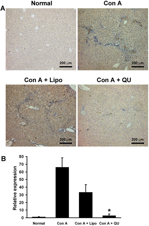
Figure 3. Quercetin inhibited the expression of NF-κB. A, Sections were immunostained for the NF-κB p65 subunit; the dark yellow or brown colors indicate NF-κB-positive cells. B, Results of real-time RT-PCR for the NF-κB p50 subunit. Data are reported as the means and standard deviation of the relative expression levels of NF-κB. QU: liposomal quercetin; lipo: free liposomes; normal: saline. *P<0.01, compared to Con A control (one-way ANOVA followed by the LSD test).