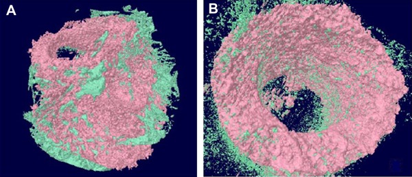
Figure 3. Micro-CT image of the magnesium scaffold: the new bone tissue in-growth 3 months post-operation (A). Micro-CT image of hydroxyapatite scaffold: visible new bone scattered on the surface of hydroxyapatite 3 months post-operation (B) (green indicates newly generated bone tissue).

