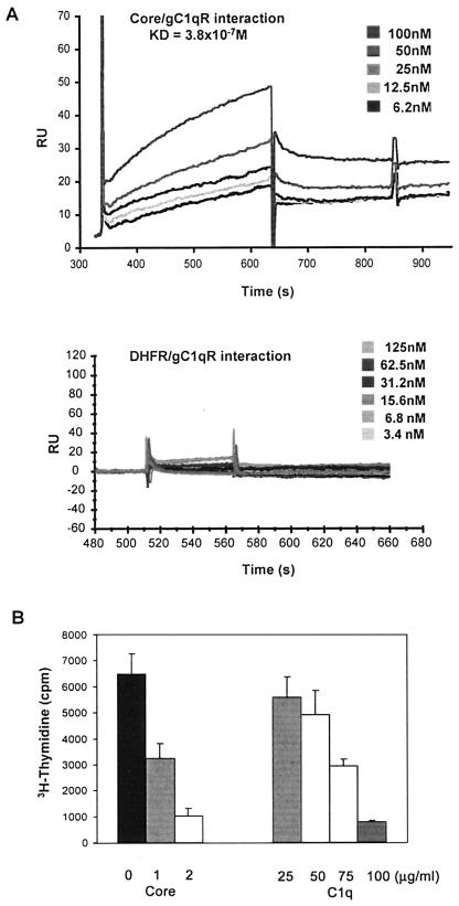
FIG. 4.
The HCV core, which binds gC1qR with an affinity similar to that of C1q, elicits a stronger inhibitory signal on T cells. (A) Kinetics of the HCV core-gC1qR interaction and its dissociation constant (Kd) determined by BIAcore analysis. Various concentrations of the HCV core or DHFR were passed over gC1qR (50 μg/ml) covalently coupled to a dextran chip at a flow rate of 50 μl/min. The Kd for the HCV core-gC1qR interaction, as determined by using the BIA analysis computer program, is shown. Results are presented as RU/s, where 1 RU = 1 pg/mm2. (B) Dose-dependent inhibition of T-cell proliferation by the HCV core or C1q. A total of 2 × 10⁵ ConA-stimulated PBMC were incubated with various concentrations of either the HCV core or C1q for 5 days at 37°C. [3H]thymidine was added to the cultures 18 h prior to harvesting, and incorporation was determined by using a MicroBeta liquid scintillation counter. Error bars indicate standard deviations. The results were reproducible in three independent experiments.

