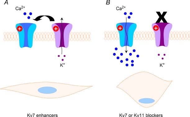
Figure 1. Schematic representation of the functional role of potassium channels in uterine smooth muscle contraction.

Left-hand panel shows that open K+ channels result in membrane hyperpolarization that indirectly limits the opening of voltage-dependent calcium channels shown in blue. This results in a less contracted smooth muscle. In the right-hand panel, the potassium channels are non-functional due to blockade, loss-of-function mutations or trafficking defects. This leads to membrane depolariziation, and the open probability of the calcium channels increases. The concomitant influx of calcium contributes to smooth muscle contraction.
