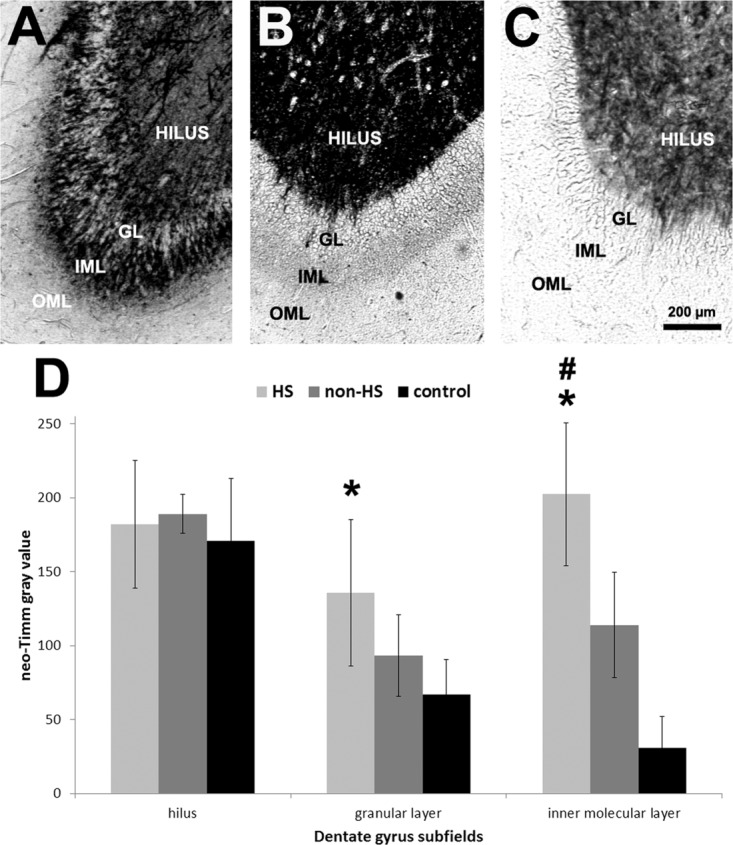
FIGURE 2.

Neo-Timm histochemistry of the human dentate gyrus. Low magnification images of neo-Timm stained DG revealed the typical pattern with dark stained hilus in HS (A, light gray bars), non-HS (B, gray bars), and controls (C, black bars) with increased mossy fiber sprouting in HS GL and IML. Gray values in the HS GL were higher than in controls and in the IML were higher in HS group when compared to non-HS and controls. Values indicated as mean ± std. deviation. Asterisk indicate significant statistical difference (P < 0.05) between epileptic and control group. Hash sign indicate significant statistical difference (P < 0.05) between epileptic groups. OML: outer molecular layer; IML: inner molecular layer; GL: granular layer.
