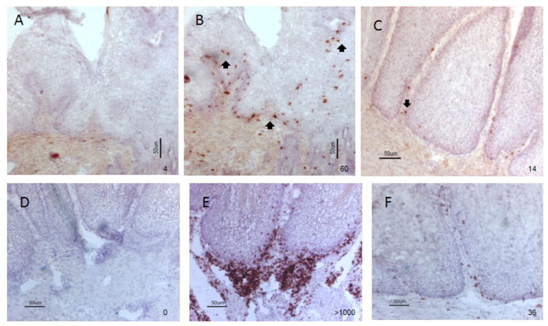
Figure 5.

Immunohistological detection of CD8+ (A-C) and CD4+ T (D-F) cells at the sites of lymphocytic infiltration. A and D) Negative control omitting monoclonal antibody against rabbit CD8 and CD4 during the staining; B and E) A population of CD8+ and CD4+ T cells (red) were found around the basal layer of the tumor (shown by arrows) that showed high levels of lymphocytic infiltration as shown in figure 4A (10×); C and F) Low numbers of CD8+ and CD4+ T cells were found in the persistent tumors that had limited lymphocytic infiltration (10×) respectively.
