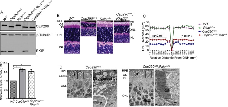
Figure 2.
Characterization of double-knockout mice. (A) Immunoblot analysis of retinal extracts (30 μg) of mice of indicated genotypes was performed using antibodies against CEP290, RKIP and β-tubulin (loading control); RKIP immunoreactive band is not detected in the Rkipko/ko lanes. Also, predicted shorter deleted variant of CEP290 is detected in Cep290rd16 retinal extract. Lower panel shows quantitative analysis of the band intensity of RKIP in the Cep290rd16 and Cep290rd16:Rkip+/ko mouse retina relative to WT. (B) Histological analysis of WT and mutant retinas of indicated genotypes was performed to assess retinal morphology. Thinning of the ONL was observed in the Cep290rd16 retina, whereas improved thickness of the ONL is detected in the Cep290rd16:Rkipko/ko retina. (C) Morphometric analysis of retinas, also showed an overall statistically significant (P < 0.01) improvement in the thickness of the ONL. Retinas from five different mice of each genotype were analyzed in this experiment. (D) TEM analysis of Cep290rd16 (left panel) and Cep290rd16:Rkipko/ko (right panel) mice was performed to assess detailed photoreceptor morphology. Arrow in upper panel points to indistinguishable OS and IS while arrow in lower panel depicts organized OS and IS. Inset shows disorganized OS in Cep290rd16 photoreceptors and correctly developed and stacked OS discs in Cep290rd16:Rkipko/ko retina. Scale bars: 5 and 1 μm (inset).

