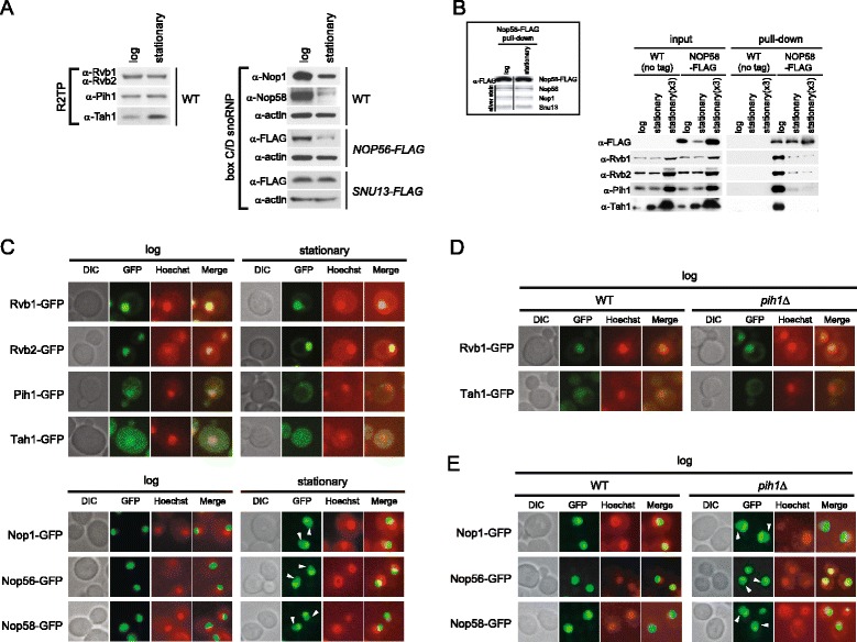
Figure 5.

The dependence of R2TP-Nop58 interaction on cell growth phase. (A) Western blot analysis of steady-state levels of box C/D snoRNP and R2TP proteins in log and stationary phase cells. (B) Western blot analysis of R2TP proteins bound to Nop58-FLAG pulldown complex in log and stationary phase cells. The pulldowns were performed using Nop58-FLAG cell lysates from equal weight of log and stationary phase cell pellets and three times by weight of stationary phase cell pellet [stationary (x3)]. As a negative control, untagged wildtype strain was used. Inset shows the Western blot or silver stain analysis of snoRNP proteins of Nop58-FLAG pulldown purified from log and stationary phase cells. (C) Subcellular localizations of endogenously GFP-tagged R2TP and box C/D snoRNP proteins in log and stationary phases. Cells were stained with Hoechst33342 and then analyzed for GFP or Hoechst33342 fluorescence. The images of DIC, GFP (green), and Hoechst33342 (red), and the merged pictures are shown. White arrowheads show nucleoplasmic GFP signals of Nop1-GFP, Nop56-GFP, and Nop58-GFP. (D) Subcellular localizations of endogenously GFP-tagged Rvb1 and Tah1 in WT and pih1Δ log phase cells. (E) Subcellular localizations of endogenously GFP-tagged Nop1, Nop56, and Nop58 in WT and pih1Δ log phase cells.
