Figure 8.

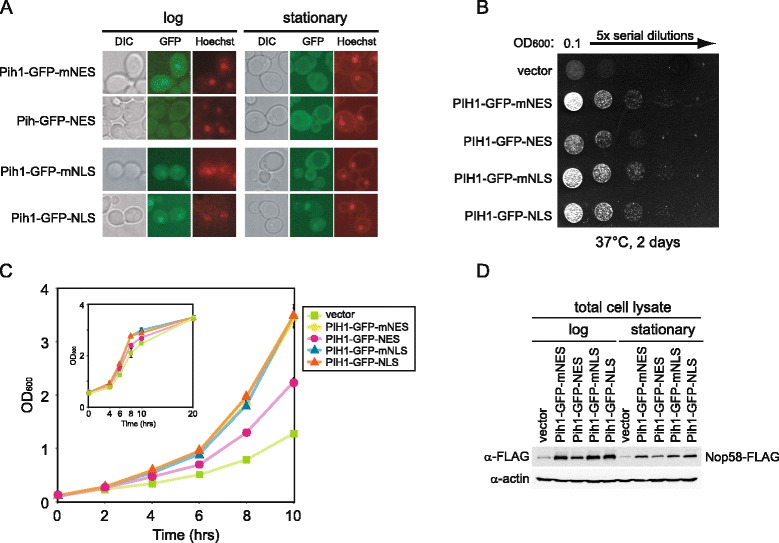
Effect of targeting Pih1 to cytoplasm or nucleus on cell growth. (A) Pih1-GFP was fused with nuclear export signal (NES), mutagenized nuclear export signal (mNES), nuclear localization signal (NLS), or mutagenized nuclear localization signal (mNLS), and expressed from pRS315 vector under the PIH1 promoter in Nop58-FLAG pih1Δ strain. Cells were stained with Hoechst33342 and then analyzed for GFP or Hoechst33342 fluorescence. The images of DIC, GFP (green), and Hoechst33342 (red) are shown. (B) Dilution spot assay of the strains described in A. Cells were serially diluted fivefold, spotted onto SD-Leu plate, and incubated for 2 days at 37°C. (C) Growth curves in liquid culture for the different indicated strains. Cells were grown in SD-Leu medium at 37°C and OD600 was measured every 2 h. Inset shows growth curve of the same strains until stationary phase. (D) Western blot analysis of total cell lysate of the different transformants. Cell lysates were prepared from cells grown at 37°C until log or stationary phase.
