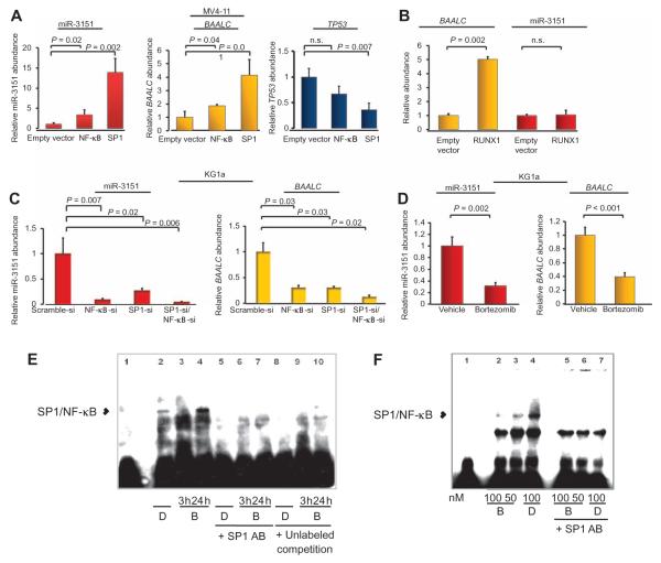
Fig. 7. Bortezomib lowers miR-3151 and BAALC expression.
(A) Effect of SP1 and NF-κB overexpression in MV4-11 cells on the abundance of mature miR-3151 (left), BAALC1,6,8 mRNA (middle), and TP53 mRNA (right). Data are normalized to the abundance in the empty vector–infected cells (mean ± SD, n = 3 biological replicates; P values were determined using two-tailed Student’s t test). (B) Effect of overexpression of RUNX1 in MV4-11 cells on the abundance of miR-3151 and BAALC mRNA (mean ± SD, n = 3 biological replicates; P values were determined using two-tailed Student’s t test). (C) Effect of knocking down SP1, NF-κB, or both in KG1a cells on the abundance of miR-3151 and BAALC mRNA (mean ± SD, n = 3 biological replicates; P values were determined using two-tailed Student’s t test). (D) KG1a cells were exposed to 100 nM bortezomib for 3 hours. The abundance of the indicated RNAs was measured by qRT-PCR and normalized to the amount in the vehicle (DMSO)–treated samples (mean ± SD, n = 3 biological replicates; P values were determined using two-tailed Student’s t test). (E) EMSA of TSS-3151 with nuclear extracts from DMSO (D)–or bortezomib (B)–treated KG1a cells. Nuclear extracts were prepared either 3 or 24 hours after DMSO or bortezomib addition in the presence or absence of 1.2 μg of SP1 antibody or unlabeled TSS-3151 in an amount equal to that of the biotin-labeled TSS-3151. (F) EMSA of TSS-BAALC. The experiment was performed with nuclear extracts (NE) of KG1a cells exposed to DMSO (D) and either 50 or 100 nM bortezomib (B) in the presence or absence of 1.2 μg of SPI antibody. Nuclear extracts were prepared 3 hours after the addition of DMSO or bortezomib. Data in (E) and (F) are representative of two independent experiments.

