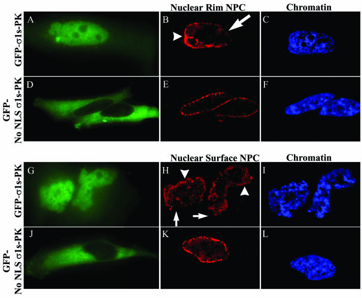
FIG. 6.
Nuclear localization of σ1s induces NPC clustering. HeLa cells were transfected with GFP-σ1s-PK (A to C and G to I) or GFP-No NLS σ1s-PK (D to F and J to L). Digital microscopy of NPC nucleoporin-labeled cells expressing GFP-σ1s-PK (A and G) showed clustering of NPCs at the surface of the nucleus (arrowheads in panel H) and at the nuclear rim (arrowheads in panel B), while some areas of the NE were depleted of NPC (arrows in panels B and H). Cells in which σ1s is expressed but is not able to translocate to the nucleus (D and J) showed conventional NPC staining (E and K). Hoechst 33342 dsDNA stain was used to define nuclei (C, F, I, and L).

