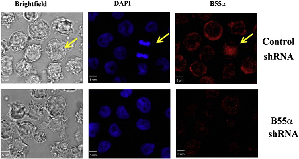
Fig. 4.
Imaging of B55α in OCI-AML3 cells by confocal immunofluorescence microscopy. B55α was imaged in OCI-AML3 cells containing control (NS) lentiviral plasmid or cells containing B55α shRNA lentiviral plasmid by confocal IF microscopy as described in the “Materials and methods”. Images are Brightfield (gray), DAPI stained nuclei (blue), and B55α stained (red).

