
Figure 4.
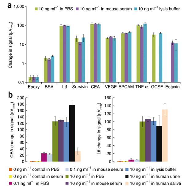
Multiplex protein detection in a diversity of media. (a) A panel of eight human tumor markers and a BSA negative control and epoxy control indicate matrix-insensitive protein detection when shifting from PBS to mouse serum to lysis buffer. No bar graph is shown for the 10 ng ml-1 GCSF spiked into mouse serum or 10 ng ml-1 of eotaxin spiked into PBS due to sensor corrosion during the experiment. The error bars represent means ± s.d. Ltf, lactoferrin. (b) Matrix-insensitive protein detection across a range of concentrations (0 ng ml-1 control, 0.1 ng ml-1 spiked samples and 10 ng ml-1 spiked samples) for CEA and Ltf in PBS, mouse serum and lysis buffer. In addition, detection of 10 ng ml-1 CEA and Ltf spiked in PBS, mouse serum, lysis buffer, human urine and human saliva is presented. The error bars represent means ± s.d.