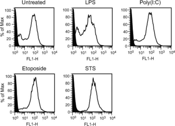
Figure 3.

Annexin V staining of microparticles. MPs released by activated, apoptotic and untreated RAW 264.7 cells were stained with FITC-annexin V. The levels of staining were measured by flow cytometry. Data shown are representative of three separate experiments. Black indicates unstained controls. As these data indicate, supernatants from all cultures had particles that bound annexin V, with particles from untreated and LPS- stimulated cells showing a larger percentage of MPs with lower levels of staining.
