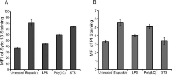
Figure 4.

Flow cytometry analysis of microparticles. MPs released from RAW 264.7 cells treated to undergo activation or apoptosis were stained with either SYTO 13 (A) or PI (B). The stained samples were then analyzed using flow cytometry. Data shown are representative of three separate experiments.
