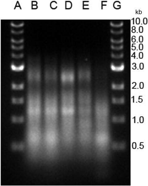
Figure 6.
Analysis of microparticle DNA by gel electrophoresis. DNA was isolated from purified MPs from RAW 264.7 cells undergoing activation or apoptosis. The samples were analyzed on a 1% agarose gel in TAE buffer at a concentration of 200 ng/lane. The samples were visualized by staining with ethidium bromide. Lanes A and G show molecular weight markers. DNA from MPs from the following cultures were analyzed: (B) untreated; (C) etoposide; (D) LPS; (E) poly (I:C) and (F) STS.

