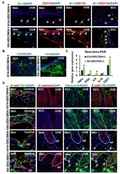
Figure 2. K6-ODC/SKH-1 & K14-ODC/SKH-1 mice show alterations in UVB-induced inflammatory responses.

(A) Immunofluorescence staining of MDSCs (CD11b & Gr-1) in UVB-irradiated skin & tumors excised from K6-ODC/SKH-1 & K14-ODC/SKH-1 mice; (B) F4/80 positive cells (macrophage surface marker) in UVB-irradiated K6-ODC/SKH-1 compared to K14-ODC/SKH-1 mice; (C) Relative gene expression of pro-inflammatory cytokines in UVB-irradiated skin of K6-ODC/SKH-1 & K14-ODC/SKH-1 mice; (D) Coimmunostaining showing expression of EMT markers, E-cadherin and fibronectin in control & UVB-induced skin and tumors. Hair follicular regions in control and UVB-irradiated skin are denoted as ‘HF’ in represented images.
