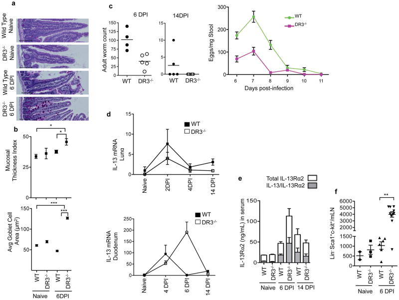
Figure 5. DR3 is not required for IL-13 production and clearance of parasites in Nippostrongylus brasiliensis infection.
(a) Representative PAS-stained duodenum sections from wild-type or DR3−/− mice without or 6 days post infection (DPI) with Nippostrongylus brasiliensis. Scale bar=100μm. (b) Mucosal thickness and goblet cell area in these samples are shown for 4–9 mice per group with each dot representing the average within a group. Data represent mean ± SEM, analyzed with Mann Whitney (*p<0.05, ***p<0.001). (c) Adult worm counts from the intestine at 6 and 14 days post infection, and counts of eggs in the stool over the course of infection. Data represent mean ± SEM. (d) IL-13 mRNA expression in the duodenum over the course of infection normalized to β2m expression and shown relative to expression levels in untreated WT mice with mean ± SEM. (e) Serum levels of IL-13Rα2 (white bars) and IL-13 bound to IL-13Rα2 (grey bars) measured in serum at the indicated times after Nippostrongylus infection in the indicated mice. (f) ILC2 quantitated in mesenteric lymph nodes of uninfected and 6DPI Nippostrongylus infected mice using Lin−(CD3CD4CD8CD11bCD19MHCIIFcεRI) ckit+ Sca1+ as markers of ILC2. See also Figures S4 and S5.

