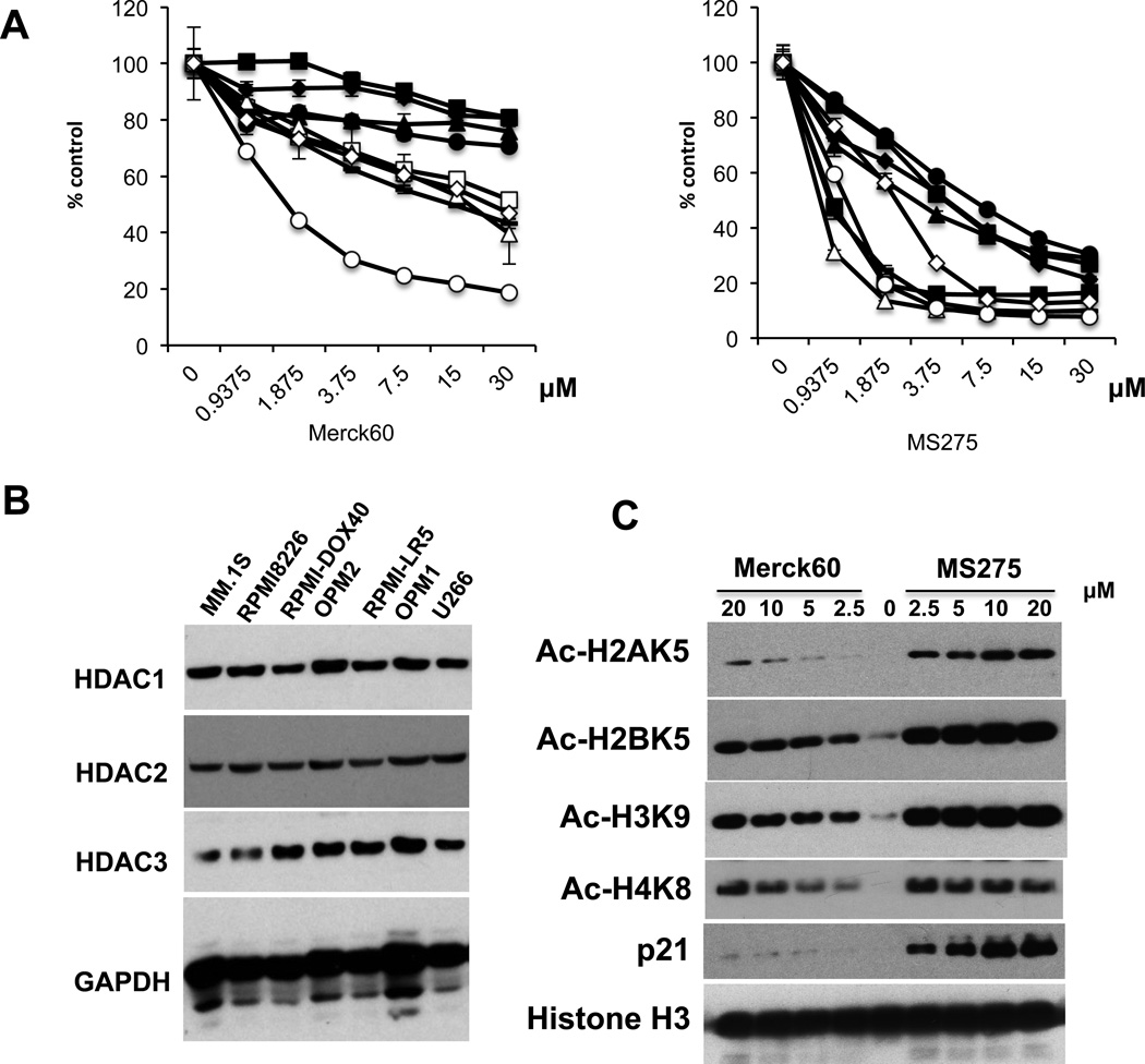
Figure 1. MS275 is more cytotoxic than Merck60 in MM cells.
(A) MM.1S (□), RPMI8226 (●), U266 (▲), H929 (■), MM.1R (∆), RPMI-LR5 (♦), OPM1 (−), OPM2 (○), RPMI-DOX40 (◊) MM cells were cultured with Merck60 (left panel) or MS275 (right panel) for 48 hours. Cell growth was assessed by MTT assay. All experiments were performed 3 times in quadruplicate. Data represent mean ± SD. (B) Whole cell lysates from MM cells lines were subjected to immunoblotting to assess HDAC1, 2 and 3 expression. GAPDH served as a loading control. (C) Whole cell lysates from RPMI8226 cells treated with Merck60 or MS275 for 12h were subjected to immunoblotting with anti-Ac-H2AK5, -Ac-H2BK5, -Ac-H3K9, -Ac-H4K8, -p21WAF1. and -histone H3 antibodies (Abs).

