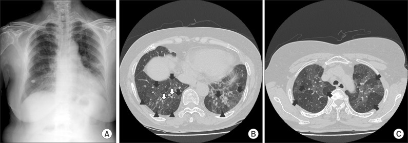
Figure 1.
(A) The chest radiograph demonstrates diffuse reticulonodular opacities in both lungs. (B) A high resolution computed tomography (HRCT) scan reveals diffuse patchy ground glass opacity (black arrows), mosaic perfusion (black arrowheads) and traction bronchiectasis (white arrows) in both lungs. (C) Air-trapping (black arrows) is noted in an expiratory view of the HRCT scan.

