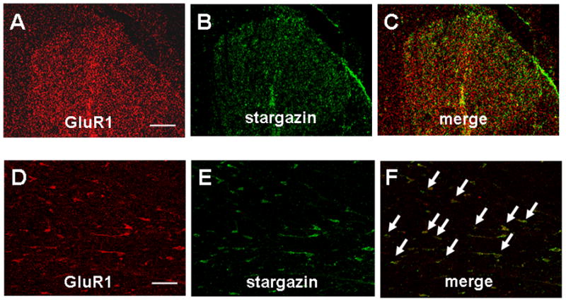
Fig. 3. Colocalization of stargazin and GluR1 in rat dorsal horn.

Representative confocal images of GluR1 (A and D, red) and stargazin (B and E, green) immunoreactivity in the dorsal horn of L5–L6 spinal segment. Colocalization (merge) is shown in yellow/orange (C and F). Double-labeled neurons are indicated by arrows. A, B, and C are shown in lower magnification. D, E, and F are shown in higher magnification. Scale bars: 100 μm (A) and 20 μm (D).
