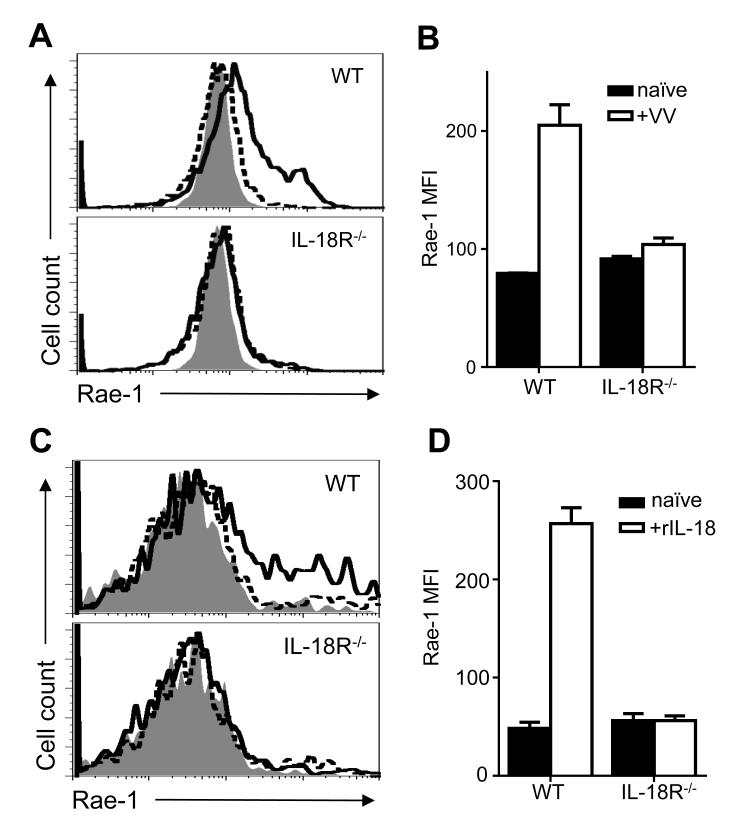
FIGURE 5.
IL-18 signaling on DCs is required for Rae-1 upregulation to VV in vivo. WT or IL-18R−/− mice were infected with 5 × 10⁶pfu VV i.p. or left uninfected (naïve). 24 h after infection, splenocytes were assayed for Rae-1 surface expression on CD11c+CD11b+B220− cDCs. (A) Histograms compare Rae-1 expression on cDCs from infected (thick line) and naïve (dotted line) WT (top panel) and IL-18R−/− (bottom panel) mice. Shaded histogram indicates isotype control. (B) The mean fluorescent intensity ±s.e.m. of Rae-1 on cDCs is shown (n=3 infected mice). Data is representative of three independent experiments. Interaction term for two-way ANOVA is p<0.01. (C) Histograms compare Rae-1 expression on WT (top panel) and IL-18R−/− (bottom panel) bone marrow-derived DCs cultured with 5 ng/mL rIL-18 (thick line) or left untreated (dotted line) for four days. Shaded histogram indicates isotype control (D) The mean fluorescent intensity ±s.e.m. of Rae-1 on bone-marrow derived DCs is shown (n=3 wells). Data is representative of three independent experiments. Interaction term for two-way ANOVA is p<0.0001.

