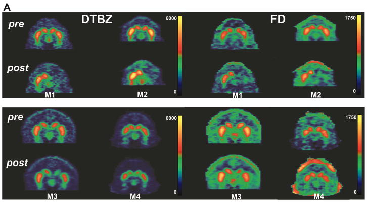
Figure 6. PET imaging of DTBZ and FD.

Representative coronal PET images are shown from two control primates (A; monkeys M1 and M2) and two C3-treated monkeys (B; monkeys M3 and M4) just prior to intracarotid MPTP (top panels) and at the end of two months of treatment with placebo or C3 (bottom panels). Images of DTBZ (left panels) and FD (right panels) are shown. Note the bilateral uptake of each tracer pre-MPTP, showing the tear-drop-shaped substantia nigra bilaterally for all four animals. However, at the end of the study, there is significantly less uptake of both tracers on the lesioned side in placebo-treated animals compared to C3-treated monkeys, or conversely, there is preservation of DTBZ and FD in C3-treated animals.
