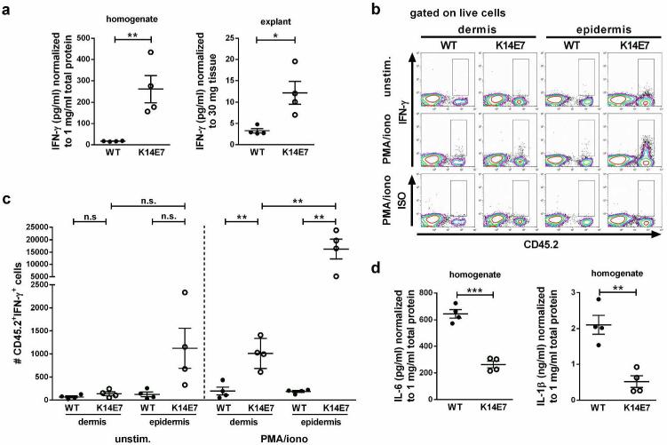
Figure 1. Elevated production of IFN-γ in K14E7 compared to wild type skin.
a IFN-γ concentrations in wild type and K14E7 skin homogenates and supernatants of skin explants (4 mice per group). Data points indicate individual mice analyzed in at least 2 independent experiments. b,c Flow cytometric analysis of cells isolated from dermis and epidermis of wild type and K14E7 mice, unstimulated or stimulated with PMA and ionomycin. b Total live cells gated for CD45.2+IFN-γ+ cells. Plots are representative of 4 independent experiments (2 mice per strain per experiment). c Numbers of CD45.2+IFN-γ+ cells in wild type and K14E7 dermis and epidermis obtained from 4 independent experiments. Bars indicate means±SEM. d IL-1β and IL-6 concentrations in wild type and K14E7 skin homogenates (4 mice per group).

