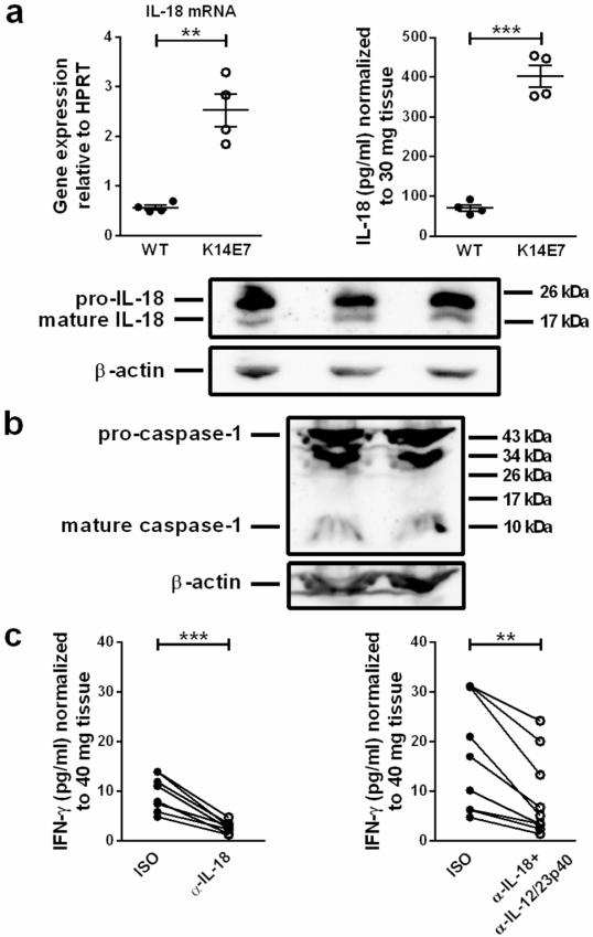
Figure 4. IL-18 production is elevated and induces IFN-γ production in K14E7 skin.
a IL-18 mRNA expression in C57BL/6 and K14E7 skin (4 mice per group). IL-18 concentrations in supernatants of C57BL/6 and K14E7 skin explants (20 h; 4 mice per group). Means±SEM are indicated. a,b Western Blots showing (a) pro-IL-18 (24 kDa), mature IL-18 (18 kDa) (n=3), (b) pro-caspase-1 (45 kDa) and mature caspase-1 (10 kDa) (n=2) in K14E7 skin lysates. β-actin served as loading control. Results are representative of at least 2 independent experiments. c IFN-γ concentrations in supernatants of K14E7 skin explants cultured with 10 μg/mL isotype or anti-IL-18 and/or anti-IL-12/23p40 antibodies for 40 h. Data are paired observations for the same mouse, analyzed in 2 independent experiments (8-9 mice per treatment).

