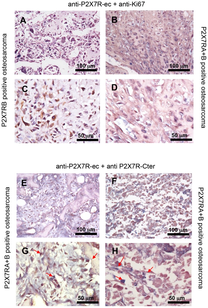
Figure 2. Double immunohistochemistry on human osteosarcomas.
Co-immunostainings of osteosarcoma tissue array either with the anti-P2X7R-ec plus anti-Ki67 antibodies (A–D), or with the two primary anti-P2X7R antibodies (E–H) were carried out as described in Materials and Methods. (A,C): representative P2X7RB positive osteosarcoma showing a higher number of Ki67 positive nuclei (stained in blue) respect to a representative P2X7RA+B tumour (B,D). (E–H): representative P2X7RA+B positive tumours demonstrating the presence of a mix of cells some (stained in brown) positive for P2X7RB only, some others (stained in blue-brown) positive for both P2X7RA and B. Two different magnifications, 20x (A,B,E,F) and 40x (C,D,G,H) are shown.

