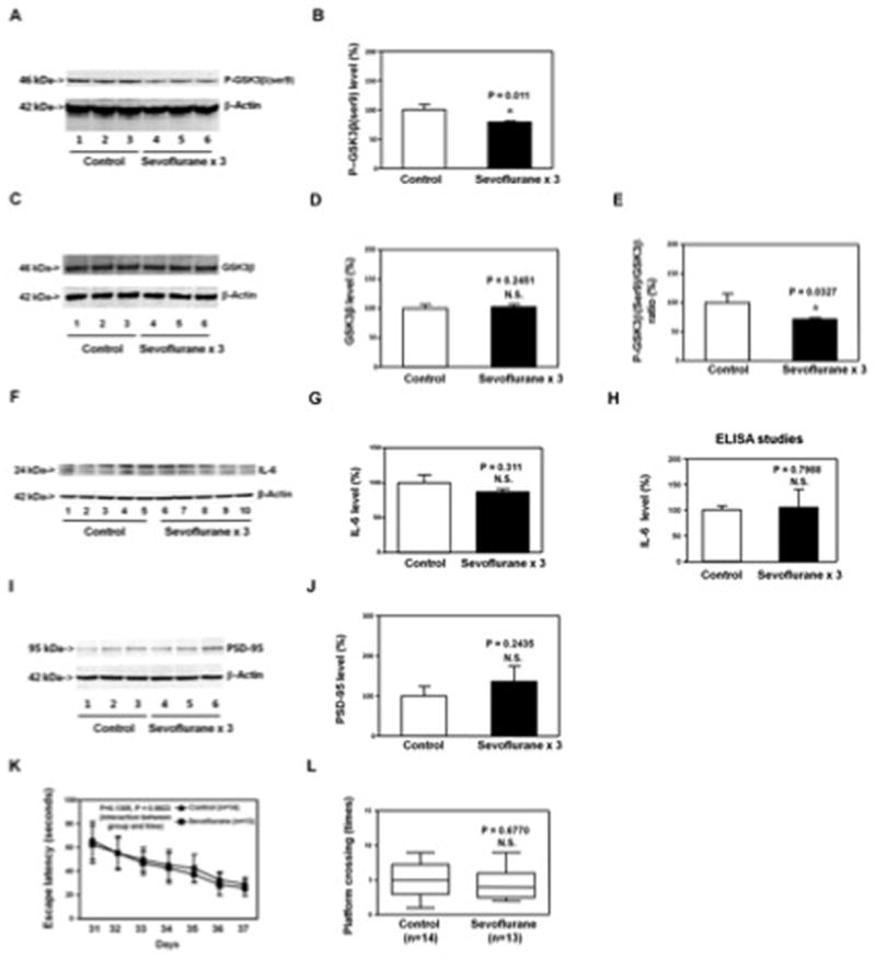
Figure 8. Anesthesia with 3% sevoflurane two hours daily for three days in P6 Tau knockout (KO) mice induces GSK3β activation. The sevoflurane anesthesia induces neither increases in IL-6 levels in hippocampus of the mice nor cognitive impairment in the mice.

A. The anesthesia with 3% sevoflurane two hours daily for three days in P6 Tau KO mice decreases P-GSK3β(ser9) levels as compared to the control condition in the hippocampus of the mice (harvested on P8). There is no significant difference in β-Actin levels in the hippocampus of the mice between the sevoflurane anesthesia and control condition. B. Quantification of the Western blot (A) shows that the sevoflurane anesthesia decreases P-GSK3β(ser9) levels as compared to control condition. C. The anesthesia with 3% sevoflurane two hours daily for three days in P6 Tau KO mice does not significantly alter GSK3β levels as compared to control condition in hippocampus of the mice (harvested on P8). There is no significant difference in β-Actin levels in the hippocampus of the mice between the sevoflurane anesthesia and control condition. D. Quantification of the Western blot (C) shows that the sevoflurane anesthesia does not significantly alter GSK3β levels as compared to control condition. E. Quantification of the Western blots (A and C) shows that the sevoflurane anesthesia decreases the ratio of P-GSK3β(ser9) to GSK3β in hippocampus of the mice. F. The sevoflurane anesthesia does not increase the IL-6 levels in the hippocampus of the Tau KO mice as compared to the control condition. There is no significant difference in β-Actin levels in hippocampus of the mice between the sevoflurane anesthesia and control condition. G. Quantification of the Western blot shows that the sevoflurane anesthesia does not increase the IL-6 levels in the hippocampus of the Tau KO mice as compared to the control condition. H. ELISA studies show that the sevoflurane anesthesia does not increase IL-6 levels in the hippocampus of the Tau KO mice (harvested on P8) as compared to the control condition. I. The sevoflurane anesthesia does not decrease the PSD-95 levels in the hippocampus of the Tau KO mice (harvested at P31) as compared to the control condition. There is no significant difference in β-Actin levels in hippocampus of the mice between the sevoflurane anesthesia and control condition. J. Quantification of the Western blot shows that the sevoflurane anesthesia does not decrease the PSD-95 levels in the hippocampus of the Tau KO mice (harvested at P31) as compared to the control condition. K. Anesthesia with 3% sevoflurane two hours daily for three days in P6 Tau KO mice dose not increase escape latency of mice swimming in the MWM as compared to the control condition (tested from P31 to P37). Two way ANOVA with repeated measurement analysis shows that there is no statistically significant interaction of treatment and group based on escape latency of MWM between mice following the control condition and mice following the sevoflurane anesthesia (3% for two hours daily for three days) in the MWM (control: N = 14, sevoflurane: N = 13). L. Anesthesia with 3% sevoflurane two hours daily for three days in the P6 Tau KO mice does not reduce the platform crossing times of mice swimming in the MWM as compared to the control condition tested at P37 (control: N = 14, sevoflurane: N = 13; P = 0.6770). P, phosphorylated; KO, knockout; IL-6, interleukin-6. PSD-95, postsynaptic density-95; GSK-3β, glycogen synthase kinase 3β; ser9, serine 9; MWM, Morris Water Maze; ANOVA, analysis of variance. N = 6 in each group (biochemistry studies); N = 13 – 14 in each group (behavioral studies).
