Fig. 7.
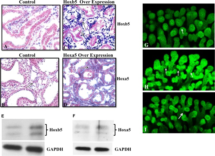
Over expression of Hoxb5 or Hoxa5 protein in fetal mouse lungs caused different changes in airway branching patterns. In control lungs (a, b), scattered Hoxb5 (a) and Hoxa5 (b) positive cell (blue staining) clusters were seen underlying the airway epithelium. However, Hoxb5 plasmid transfected lungs (c) had intense and diffuse staining (arrows in c) surrounding airways with narrow lumens and apparent multiple branch points. Hoxa5 transfected lungs (d) had diffuse mesenchyme expression of Hoxa5 (arrows in d) surrounding developing airways with wider lumens than the Hoxb5 transfected lungs. e, f Western blot confirms increased protein levels when lungs were transfected with either the Hoxb5 (e) or Hoxa5 (f) expressing plasmid. g, h, i E-Cadherin whole mount confocal images show that compared to control lungs (g), Hoxb5 transfected lungs (h) had multipodal and 3D airway structures (arrows in h). Hoxa5 transfected lungs (i) developed a more finely arborized airway branching pattern (arrows in i) that appeared better organized than that seen with Hoxb5 over expression. 20× Mag, N = 3 lungs per condition

