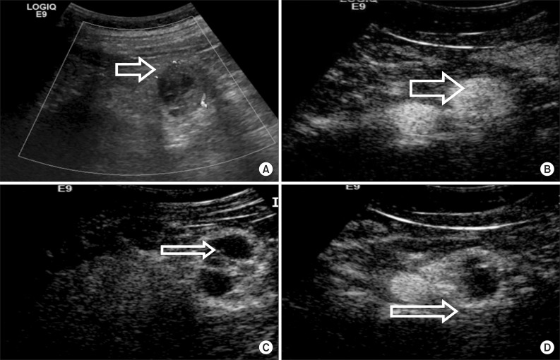
FIG. 1.
A 42-year-old man with clear cell renal carcinoma. (A) Conventional ultrasonography demonstrated that a hypoechogenic mass with a diameter of 2.8 cm was located in the left lower kidney. (B) Contrast-enhanced ultrasound (CEUS) imaging of the arterial phase showed a diffuse, heterogeneous enhanced tumor. (C) CEUS imaging at the late phase showed washout of the tumor. (D) CEUS imaging at the late phase showed perilesional enhancement with slow centripetal fill in a rim-like pattern.

